Pulsed-wave tissue Doppler at the tricuspid level of the lateral right


Tissue doppler imaging

A) An oesophagogastroduodenoscopy 6 weeks postoperative showing tumour
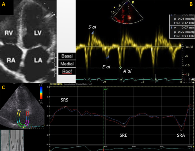
Tissue Doppler Imaging and strain rate of the left atrial lateral

Tissue Doppler systolic annular velocity and myocardial performance index as predictors of right ventricular affection and culprit lesion location in acute inferior myocardial infarction - ScienceDirect

Pulsed wave tissue Doppler imaging of the lateral tricuspid annulus

PDF) Tissue Doppler systolic annular velocity and myocardial performance index as predictors of right ventricular affection and culprit lesion location in acute inferior myocardial infarction

A Clinician's Guide to Tissue Doppler Imaging

A,B: Axial sections, portal phase of abdominal computed tomography

Pulsed-wave tissue Doppler imaging at the lateral tricuspid

Prognostic implications of impaired longitudinal left ventricular
JCM, Free Full-Text

Pulsed-wave tissue Doppler at the tricuspid level of the lateral right

Serdar BOZYEL, Assistant Professor, Assistant Professor, Cardiology

A,B: Axial sections, portal phase of abdominal computed tomography

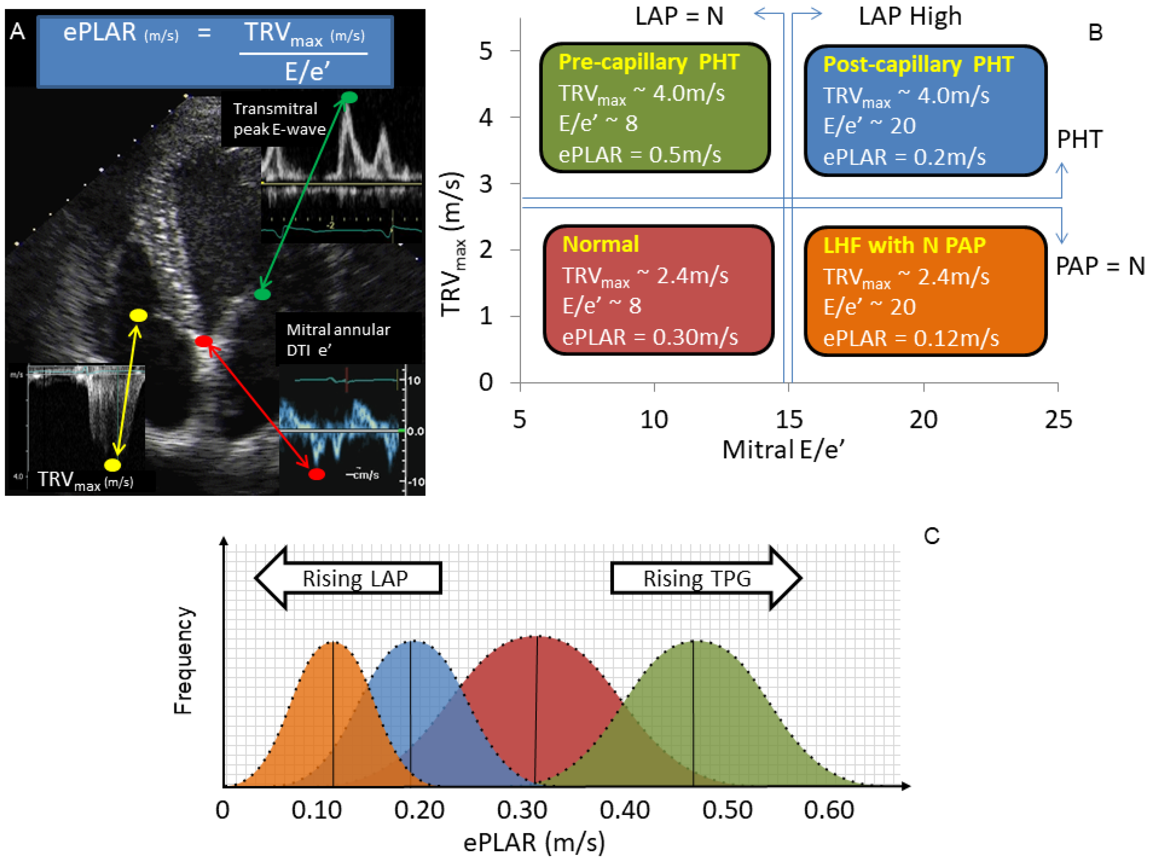
RV E/Ea ratio is 3.5 in a healthy 21-year-old subject (upper panel